You can see everything … except the data! Technical due diligence is a *must*.

You can see everything … except the data!  Technical due diligence is a *must*.
Holmes & Evans (source: NY Times)

I saw “You can see everything” with my wife on our date night last Friday. We both really liked it, and were puzzling away on the drive home about who said what to whom, who we thought was likely lying or truthful, what Elizabeth Holmes’ partner Bill Evans was really up to, whether Nathan Fielder (the journalist) was going to lunge and kiss Holmes (former CEO of Theranos) when she was hitting him with the puppy dog eyes, etc. We both knew of the events regarding Theranos generally, but were not well acquainted with the particulars prior to the movie, which probably made for better entertainment. 🙂

If you haven’t seen it yet, the movie is a meta-documentary that did a great job of immersing you in Holmes’ and Evans’ personal circumstances with their two young kids in the weeks prior to her having to report to prison in Texas in 2023 for defrauding investors out of hundreds of millions of dollars. It delves into Holmes’ and Evans’ claims of her innocence regarding her over-reaching/fraudulent statements about the company’s microfluidic blood analysis technology and its capabilities. SPOILER ALERT!! Fielder focussed on whether Holmes was intentionally deceiving people (i.e. investors, the public) when, in years past, she said things to the effect of ‘the product can do a multitude of tests simultaneously’. He was trying to determine whether she genuinely didn’t understand that this phrase would be interpreted by most to mean “the product has already successfully and conclusively demonstrated its ability to do a multitude of tests …”, not “the product can, in theory and once fully developed, do any and all of a multitude of tests …”. To get a better read on her interpretation of her own words, he gets her interpretation of the “Who’s on First” skit by Abbott and Costello, where her answers suggest that, by virtue of her very literal interpretation, she misses the double ententes that are the essence of the humour. They touch on whether she is neuro-divergent, and leave the audience to draw their own conclusions.
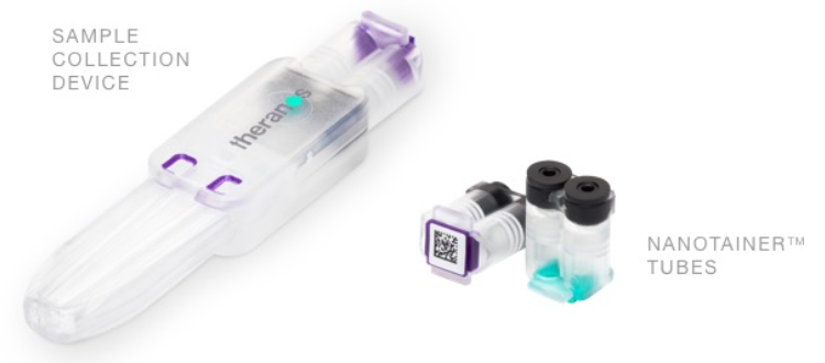
Theranos finger prick blood sampling device & sample tubes (source: Theranos.com via Wayback Machine)

The next morning, my wife and I were still sorting out all the loose ends during our dog walk. We discussed who we now believed to be truthful, and my wife was asking me about the microfluidics, analytical chemistry and business aspects of the story. This is also where my thoughts had led to, and is the reason I’m writing this article.

The short version of this post’s analysis can be summarised as follows: there’s no way the Theranos calamity would have happened had robust technical due diligence been performed. It can be put even more succinctly using the Russian proverb that the late President Reagan was fond of: trust, but verify.

As a microfluidics consultant who a) supports technology development projects, and b) performs technical due diligence investigations on behalf of prospective investors, I can tell you that I was dumbfounded as to how this situation could have arisen in the first place. While the trustworthiness of the leadership team at Theranos (or any company) would have been crucial to investor decision-making, so too should have been the imperative for, and results from, a rigorous technical due diligence investigation. My astonishment lead to a deeper dive into the events surrounding the rise and fall of Theranos, and especially into investment rounds that totalled over $900M USD from venture capitalists (VCs) and companies like Safeway and Walgreens.
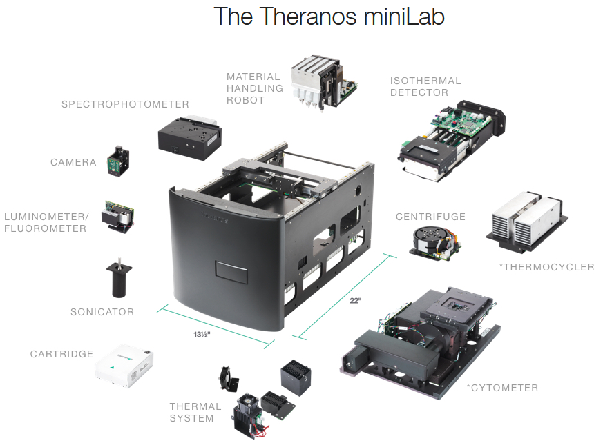
Theranos miniLab instrument (source: Theranos.com via Wayback Machine)

It turns out I’m very late to the party; much has been written on this very point. If you’re interested in the business and investment angle of the story, there is a pretty good Wikipedia entry for Theranos. and below are some references that explore various aspects of this story, including legal references, investigative journalism, an on-site interview video, and retrospective analyses and opinions on problems and failure modes.

  1. Wall Street Journal exposé on Thereanos’ inconsistent product performance, secrecy and fraudulent practices that kicked off the firestorm of trouble for the company (John Carreyrou, October 15th, 2015):
  2. Wall Street Journal exposé on Walgreens’ investment in Thereanos despite its misgivings due to secrecy regarding product performance (Christopher Weaver and John Carreyrou, May 25th, 2016):
    1. https://www.wsj.com/articles/craving-growth-walgreens-dismissed-its-doubts-about-theranos-1464207285
  3. CNN video tour of Theranos and interview with Holmes (Sanjay Gupta, August 1st, 2016):
  4. Editorial in the European Journal of Clinical Investigations criticising stealth (i.e. unpublished) research by start-up ‘unicorns’ (VC backed with valuations over USD $1B) (Ioana Cristea et al., April 1st, 2019):
  5. Editorial post mortem in the Journal of Medical Systems regarding the rise and fall of Theranos, highlighting steps such as encouraging engagement in academic collaborations, publication of scientific results, and revitalised investor due diligence (Rishub Das et al., April 4th, 2022)
  6. Lexology blog post regarding the importance of due diligence in light of Holmes’ fraud conviction (Ellen Lambrix, Bristows LLP, January 5th, 2022)
    1. https://www.lexology.com/library/detail.aspx?g=26af5d28-3c11-4440-880f-8806df327529
  7. Vaultinum blog post regarding Theranos’ secretive practices, unproven technology, and the importance of technical due diligence as an inoculant against future such travesties (Marine Yborra, May 18th, 2022)
  8. Legal summary for U.S. v. Elizabeth Holmes, et al., provided by the US Attorney’s Office, Norther California.

The answer to my question about how this could ever have come to be is essentially two-fold. Several top VC inventor firms refused to get involved when their attempts to perform technical due diligence were thwarted by Theranos’ insistence on secrecy, which they claimed was based on their need to protect their intellectual property (see reference 7). Other investors either didn’t bother trying to look that deeply, or weren’t willing to push Theranos to either a) expose its technology to scrutiny, or b) at least demonstrate product performance through independent testing against gold standard analytical methods. Walgreens investigative consultants were denied access and reported their reservations, but Walgreens persevered (see reference 2).

In the investment due diligence investigations I have performed, the start-ups have confidentially revealed all of their performance data, reports, and intellectual property (IP), and they have fully answered my innumerable questions about all aspects of their microfluidic devices, analytical instrumentation, protocols and procedures, and the performance achieved. This has allowed me in each case to write a detailed and accurate report for my client—the prospective investor—that fully described the extent to which IP, technology, performance, and the leadership team were evaluated, the findings in each of these areas, and my analysis and recommendations based on those findings. Some start-ups get rosier recommendations than others, naturally, but I have had full disclosure in all cases. Had I been met with resistance to supply information, I would have questioned the target company as to why, and then I would have clearly highlighted the resistance, missing information, and associated enormous risks in the report.

Thorough technical due diligence for investment in a deep tech-based company like Theranos should be regarded as mandatory by investors, not optional. I should note that I have not dealt with unicorn start-ups with their very aggressive growth curves, valuations, and leadership teams that are secretive about their technology. While I understand that a unicorn investment target represents a significant paradigm shift compared to a ‘normal’ start-up, and that a change in approach might be required for a technical due diligence investigation, the fundamental requirement of verifying product technology and performance must still be met. There really are no valid explanations or excuses that justify a lack of disclosure, or demonstration of performance; asking an investor to ‘take their word for it’ doesn’t pass muster. Suitable non-disclosure agreements (NDAs) can be used to safeguard the target company’s IP when revealed to the prospective investor or their consultant. Failing that, and at a minimum, performance comparisons against gold standard chemical analysis methods can still be demonstrated, even it the technology in question is treated as a secret black box to protect IP.

A lack of published results underpinning a technology does not prove the technology is faulty, but is a red flag, in my view. The increasing tendency of technology-based start-up unicorns to bypass the rigours of publishing their work in refereed journals is documented in references 4 and 5. Reluctance to publish is understandable in the context of a start-up with fast-approaching milestones and limited resources, as it is admittedly laborious and provides some level of potentially undesirable visibility into the nature of their technology. On the other hand, the process of publishing one’s work requires that the intended level of performance be obvious, robust and reproducible, which is in the interests of both the start-up and investor, and has been a successful approach for many start-up unicorns in the last decades.

My advice to investors? Review this cautionary tale about Theranos, and understand that the primary failure mode that led to the loss of mountains of money was not the fraud committed by Theranos’ leadership to cloak the inadequate performance of their product (horrible as that was), but was largely the failure on the part of investors to insist that technical due diligence be performed before the money flowed. Proper due diligence will expose whether performance results are excellent, modest, poor, or hidden, and enables the investor take appropriate action. In the case of hidden results, I recommend not investing!

Coacervates + Diseases + Microfluidics — Oh my!

Coacervates + Diseases + Microfluidics — Oh my!

I love learning about the chemistry of diseases, and about how microfluidics can be used to, firstly, better understand how they operate and, secondly, evaluate and refine potential therapeutic approaches that may become the foundation of a cure. To my delight, researchers from the Reches group at The Hebrew University of Jerusalem just published as an article entitled “Tailoring Peptide Coacervates for Advanced Biotechnological Applications: Enhancing Control, Encapsulation, and Antioxidant Properties” in ACS Applied Materials and Interfaces that looked at characterising the behaviour of coacervates (a form of chemical phase separation), how coacervates are part of many biolgical processes as well as a variety of neurodegenerative diseases (such as Alzheimer’s, Parkinson’s and Huntington’s diseases), and how their progress with coacervates could potentially apply to a variety of applications such as drug delivery.

Figure i: Liquid-liquid phase separations likely play key roles in many neurodegenerative diseases, cancers and infectious diseases. (Image from Figure 3 in Alberti et al.; © 2019 by Annual Reviews)

So, what in blazes is a “coacervate”, and how does any of this relate to understanding and curing diseases, you ask? Great questions, the search for the answers to which led me to an excellent 2019 review in Annual Reviews entitled “Liquid–Liquid Phase Separation in Disease” by Alberti and Dormann at the Technical University of Dresden and Ludwig-Maximilians University in Munich, respectively. Since this part is really interesting and provides context for the relevance of the Israeli research, we’ll start here. Fair warning: this is rather complicated, but I’ll try to simplify it as much as possible, without misrepresenting anything (apologies to those in the field who detect an unwitting gaffe :-).

Liquid-liquid phase separation (LLPS) is a process by which one or several solutes in a solution self-concentrate into their own phase, essentially forming microdroplets termed condensates or coacervates. It turns out that LLPS is now believed to be fairly widespread for a number of different peptides and proteins within cells, and to underlie several biological processes; in some cases, these coacervates are essentially membraneless organelles in the cell. The review illustrates the general mechanisms by which abnormal LLPS may cause disease, such as a) genetic changes in a protein that change its solubility and thus tendency to form a coacervate, b) genetic changes in a regulator that changes when or where the coacervates forms (i.e. in the cytoplasm vs. nucleoplasm), and c) changes in physiological conditions (e.g. pH, salinity, osmotic pressure, etc.) that may promote or inhibit coacervate formation. The authors then provide generalised and specific examples of these mechanisms in action for several neurodegenerative diseases (such as Alzheimer’s, Parkinson’s and Huntington’s disease, frontotemporal dementia (FTD), amyotrophic lateral sclerosis (ALS or Lou Gehrig’s disease)), cancers (apparently many or all cancers via certain common processes such as high cellular replication) and infectious diseases (such as a large number of viruses via the formation of their coacervate viral factories, as well as many bacterial and fungal infections via common processes such as solidified cytoplasm in drug-resistant strains). Their Figure 3, reproduced above right as Figure i, is a helpful illustration of these processes happening in a cell.

Specific examples of these mechanisms are beyond the scope of this post (… and beyond my understanding of the biochemistry, if I’m being honest :-), but I find even the generalised examples fascinating, and maybe you will too. Paraphrasing directly from their caption for the figure above, here are some. In neurodegenerative diseases, mutations, abnormal posttranslational modifications (PTMs, normally used to complete the synthesis of a protein), altered subcellular localization (i.e. getting the proteins to the wrong part of the cell), or impaired protein quality control (PQC; an enzyme system that might e.g. keep a transported protein from phase-separating and losing function) can promote the formation of ectopic condensates (phase separation in the wrong cellular location) as well as precipitating to a solid, both leading to protein aggregates in diseased tissue. These aggregates can impair ribonucleoprotein (RNP) granules and cellular factors (proteins), contributing to neuronal dysfunction and cell death. In cancer, mutations in signaling receptors (e.g. nicotinic acetylcholine receptor, used to contract muscles in response to acetylcholine, or release dopamine in response to nicotine) can alter the formation of signalling clusters at sites of DNA transcription (into RNA) or DNA damage repair. This, in turn, can alter cellular signaling cascades, impair transcription or DNA damage repair, and thus promote a proliferative and malignant state of a cell (tumour growth). In viral infections, liquid-like viral factories form through phase separation of certain viral proteins, promoting viral genome replication or altering the antiviral immune response. Some antiviral sensors in our immune system detect “pathogen-associated molecular patterns” (PAMPs) and form a separate phase through LLPS upon binding to foreign DNA or RNA, thus stimulating an immune response.

So, why do any of these LLPS-based mechanisms matter? Well, the better they are understood, the better drugs and other therapeutic strategies can be designed to intercede and either hinder a diseases-based LLPS process, or promote/defend a natural one, to inhibit disease progress in the end. If we consider neurodegenerative diseases for example, it has been known for some time that they all show accumulations of cytoplasmic or nuclear proteins in certain regions of the brain, and that these protein aggregations are thought to drive the degeneration. The mechanisms by which this happens are fascinating, and are based on liquid-liquid phase separation.

Figure ii: Cell functions performed by the Fused in Sarcoma, or FUS, protein. (Image from Figure 3 in Dormann et al.; © 2013 by Elsevier Inc.)

In the case of ALS and frontotemporal dimentia (FTD), a key protein is “Fused in Sarcoma”, or FUS. FUS is involved in DNA transcription, repair and splicing as well as mRNA transport in cells such as neurons; in a 2013 paper entitled “Fused in sarcoma (FUS): An oncogene goes awry in neurodegeneration” published in Molecular and Cellular Neuroscience by Dormann, FUS’s role in neurodegeneration is described in detail, and its normal functions in the cell are illustrated in Figure 3 from the paper, reproduced at right as Figure ii. One failure mode is not fully performing the PTM of methylation of the arginine amino acids in the FUS protein. In FTD patients, the arginines are either unmethylated or monomethylated, which causes phase separation of the FUS, impairing its ability to act. A second failure mode involves mislocalisation of the FUS protein.

In the case of ALS, a mutation in FUS’s “nuclear localisation sequence” (NLS, the amino acid sequence ‘code’ along a protein like FUS that allows it to be imported into the cellular nucleus) impedes its ability to bind to nuclear import receptor Transportin (like an ‘admission pass’, activated by the NLS code, that allows the bearer (FUS) to enter the nucleus). Transportin not only clears its cargo protein (FUS or others) for entry into the nucleus, it also suppresses LLPS and aggregation, so impeded FUS-transportin binding leads to accumulation, phase separation and precipitation of the FUS in the cytoplasm (termed mislocalisation). In the case of FTD patients, the issue is aggregation of the Transportin itself, rendering it inactive, i.e. unable to bind to the FUS and chaperone it to the nucleus, leading again to FUS mislocalisation. Critically, these LLPS mechanisms suggest research and development opportunities in e.g. drug therapies that may block or counteract a particular disease mechanism such as genetic mutation, mislocalisation, etc.

These disease examples are the tip of the iceberg in this field of research; many more specific examples are laid out in Alberti and Dormann’s review. If the (hopefully accurate) summaries above piqued your interest, reading the full review will certainly provide a more fulsome understanding.

So, the relevance of LLPS chemistry to the modus operandi of neurodegenerative, cancer and viral diseases seems sound, but what benefit can microfluidic devices and processes confer? The answer to this is provided in the research article from Reches’ group. In a nutshell, they did a fairly thorough job of characterising synthetic coacervates made from short polypeptides, and looking at their ability to encapsulate and protect molecules within (e.g. for drug delivery).

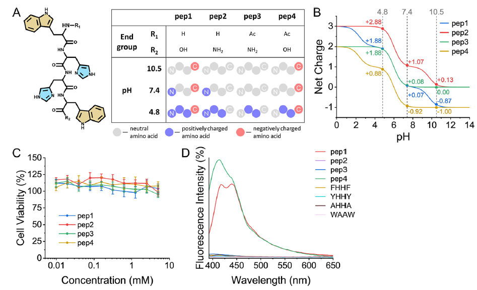
Figure iii: Four tetrapeptides used to create LLPS coacervates, together with their charges at different pH and biocompatibility. (Image from Figure 1 in Reches et al.; © 2025 by the authors and the American Chemical Society)

The researchers designed their four synthetic coacervates as being made from four short tetrapeptides, made from the amino acids tryptophan-(histidine)2-tryptophan, with different hydroxy, amino and acetyl terminations differentiating them. Their structure, charge states and biocompatibility were clearly shown in Figure 1 of their paper, reproduced at right as Figure iii. As a result of these different terminal groups, the four tetrapeptides, depicted in A, have significantly different pKa values at their acidic/basic groups, lending them different charges at a given pH, as shown in B. All four peptides performed well in ovarian cell viability tests (as a measure of biocompatibility), as shown in C.

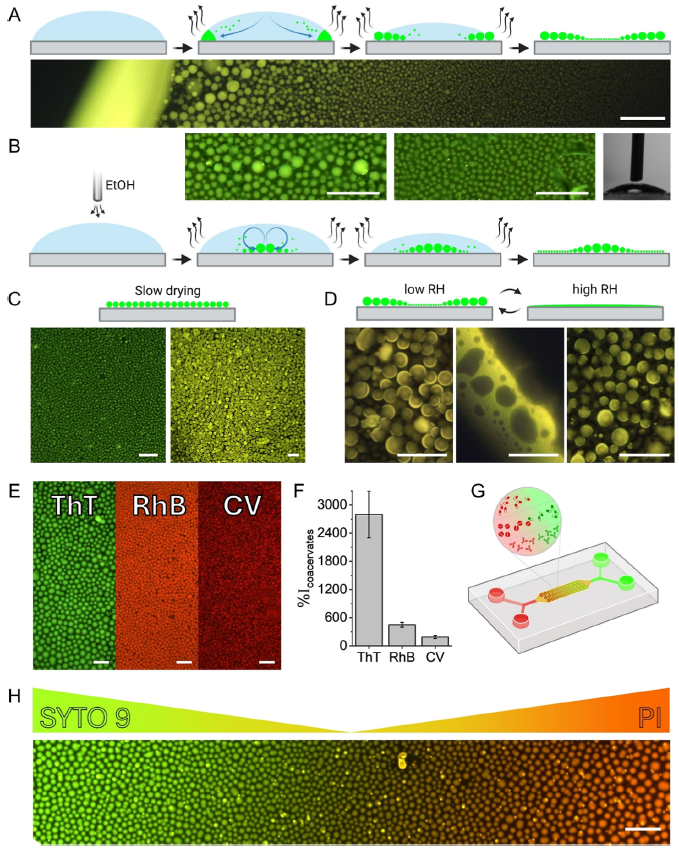
Figure iv: Formation of liquid tetrapeptide coacervates (A-D), dye encapsulation (E-F) and microfluidic generation of dye gradient (G-H). (Image from Figure 6 in Reches et al.; © 2025 by the authors and the American Chemical Society)

Characterisation of the formation and deployment of the peptide coacervate separated phase is well illustrated in Figure 6 of their paper, reproduced at right as Figure iv. In A, we see the normal evaporative formation of coacervate spheres (green) in a pH buffer solution; the smaller coacervate spheres are drawn to the solution drop perimeter by capillary action where they coalesce into larger droplets. The photo shows a fluorescence photomicrograph at the edge of the drop, with larger coacervate spheres towards the perimeter. The same process occurs in B, but ethanol vapour is blown over top of the solution drop to reduce the surface tension and in turn counter the capillary flow via the Marangoni effect (e.g. tears of wine), reversing the size distribution in the coacervate spheres vs. A. In C, slow drying is seen to produce a single coacervate size (or tight distribution, while in D, high relative humidity is shown to disassemble or merge the coacervate spheres (reversible with low RH). Frame E shows encapsulation of three dyes, thioflavin T, rhodamine B and crystal violet, in the coacervate spheres, while frame F shows the increase in fluorescence intensity for dye buffer solutions with the coacervate peptide vs. without (from ~2x to ~28x), showing the effective partitioning of the dyes in the coacervate phase. Frame G shows a schematic of the microfluidic device used to create dye gradients with green Syto 9 and red propidium iodide dyes, while frame H shows a photomicrograph of dye concentration gradient in the encapsulated dyes created by manipulation in the device. A somewhat closer look by the group at quantitative dye encapsulation showed a pH dependence, which is not surprising given the multiple acid groups and pKa values involved for each of the four tetrapeptide coacervates (referred to in Figure iii).

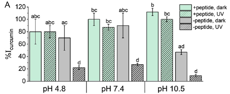
Figure v: Antioxidant properties of tetrapeptide #4 coacervate on curcumin against alkalinity and UV. (Image from Figure 7 in Reches et al.; © 2025 by the authors and the American Chemical Society)

A last important area of performance they characterised was the protective antioxidant properties of the peptide coacervates on their encapsulated charge. They chose the popular dietary supplement curcumin — the natural polyphenol in turmeric giving it its yellow colour — as the encapsulated subject to test one of the coacervate’s protective abilities. Both alkaline pH and UV irradiation cause curcumin degradation and decomposition, so optical absorbance of intact curcumin was used to measure antioxidant protection afforded by the coacervate phase in comparison to a solution with no coacervate. The results are shown in their Figure 7A, shown at right as Figure v. At an acidic pH of 4.8 or neutral pH of 7.4, the unprotected curcumin (grey) was not significantly worse than the coacervate-protected curcumin (green), though the UV radiation did significantly degrade the unprotected curcumin (grey, hatched), but not the protected curcumin (green, hatched). At the alkaline pH of 10.5, both the alkalinity and UV radiation significantly degraded the unprotected curcumin (grey & grey hatched), but the coacervate was an effective protective sheath for the curcumin (green & green hatched).

If you’ve followed this far, then congratulations are in order 😉 , and let’s now consider what all this research might eventually translate into! First, let’s list some of the salient potential benefits of the synthetic tetrapeptide coacervates in the second paper:

  • they’re made from amino acids, and are biocompatible in ovarian cells;
  • they effectively encapsulate and concentrate the dyes tested;
  • they confer alkaline and UV antioxidant protection;
  • their sphere formation morphology can be tailored by several factors including humidity, ionic strength, presence of solvents (and perhaps surfactants?); and
  • while these first peptide coacervates are a starting point, the nature of the coacervate phase could easily be tailored through choice of peptides (or other molecules or polymers), chain length, derivatisation, etc. to optimise host/drug compatibility, affinity or selectivity, encapsulation protection, etc.

How could this knowledge be built on? The authors speculated that the coacervate characterisation and performance shown in this study could lead to bio-applications such as the development biosensors as well as the encapsulated delivery of drugs and dietary supplements. I would add that there could potentially be organ-on-chip (OOC) applications, where the interactions of synthetic coacervates with the cells of a given target disease tissue, and perhaps with the natural coacervates within, could be tested. In some cases, such OOC studies would be precursors to the development of therapeutic drug delivery systems.

The authors made an initial foray into microfluidic manipulations to generate dye gradients. It is tempting to look further at the use of microfluidics to validate the effectiveness of coacervate constituents (peptides or others) to encapsulate a drug (or other ‘cargo’ molecule) under appropriate conditions, or the effectiveness of a coacervate-drug package to be delivered to the target organ tissue cells under the physiological conditions appropriate for that organ. Just as the dye gradient was shown here, other biologically relevant gradients could be explored. For coacervate encapsulation, perhaps pH, ionic strength, coacervate and drug concentrations, etc. would be of interest; for drug delivery, perhaps drug dosage, variations in physiological conditions owing to the condition itself, or owing to an additional condition — physiological changes at the cellular level in an Alzheimer’s patient that also has high cholesterol or diabetes, for example (I’m just musing about what particulars would be relevant). The ability to easily create gradients, isolate test areas, plumb reagents in and out, multiplex the conditions explored and tightly control processes on a small scale in microfluidics can help accelerate the development of a disease therapy built on the foundation of this knowledge.