Microfluidic Brain-on-a-Chip: testbed for stroke damage, therapies

Microfluidic Brain-on-a-Chip: testbed for stroke damage, therapies
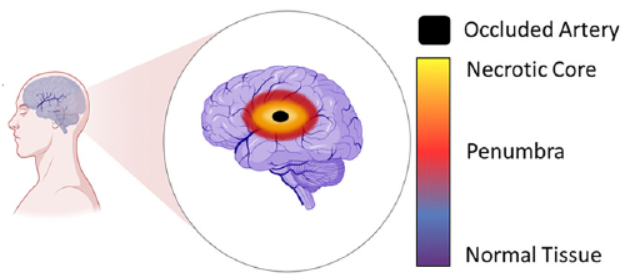
Figure 1: Schematic of brain tissue damaged from a stroke (Image © 2023 The Royal Society of Chemistry)

New research published in Lab on a Chip (September 21st) by researchers from the Ayuso lab (University of Wisconsin) and the Euan MacDonald Centre (University of Edinburgh) is an elegant example of a microfluidic organ-on-a-chip device. In this case, the organ is the brain, and the target disease is stroke. Stroke, or “brain attack”, is the brain equivalent of a heart attack. The majority of strokes are ischemic, typically caused by blood vessel blockage that leads to cell death in the brain tissue area served by that vessel.

The two research groups showed impressive results using rat astrocyte brain cells (which are a kind of “glia” — cells that support the brain’s neurons) to model the damage resulting from ischemic strokes, the response to reperfusion of nutrients to the deprived cells, and the viability of tissue grafting in an area damaged by stroke.
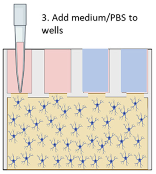
Figure 2: Cross-section of microfluidic device. (Image © 2023 The Royal Society of Chemistry)

Their microfluidic brain-on-a-chip device was made from a 384-well microtiter plate with various bottom chamber configurations machined out via CNC milling, and sealed with a polystyrene laminate using double-sided tape or solvent-assisted bonding. The chambers were filled with a collagen gel/astrocyte suspension (a proxy to mimic brain tissue) through the upper well openings, and then covered by filling the wells with either a nourishing medium, to simulate normal conditions, or a simple phosphate/borate pH buffer solution (PBS) lacking nutrients, to simulate deprivation during a stroke; see Figure 2 above.

Experiments were performed to show that their simulated normal and stroke conditions over different periods of time were reflected in terms of cell viability. In addition, the extent to which cells deprived of nutrients and oxygen during a simulated stroke were able to recover when replenished with nourishing medium was explored. The schematic shown below at left in Figure 3 illustrates a simple stroke simulation experiment (E), while the microscope image at right (F) and below (G) show the resultant effects on the astrocyte cells. Live cells are stained with with a green fluorescent dye (Cell Tracker Green), and dead cells with a red dye (propidium iodide). Magnified views of the cells directly beneath, nearby, or far from the nourishing medium are also shown (G); the further from the medium, the higher the proportion of dead cells.
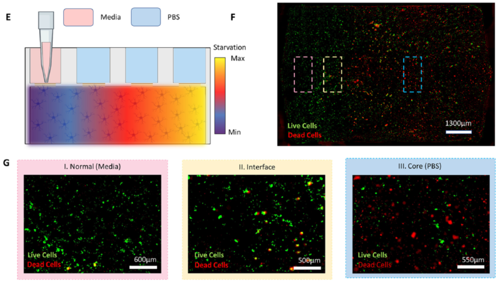
Figure 3: Stroke simulation. Nutrient gradient created with medium and pH buffer (E); resulting zones of live and dead astrocyte cells (green and red, resp.), roughly correlating to their proximity to the nourishing medium (F & close-ups in G). (Image © 2023 The Royal Society of Chemistry)

Adding different labelling and assaying approaches to the microscopy techniques above, they were able to model several other key stroke features. They demonstrated that reperfusion of the damaged pseudo-tissue area with nutrients from the medium after the simulated stroke did not reverse the damage done, replicating what has been observed clinically. Stroke damage was also evident in the deprived and reperfused cells vs. healthy cells by the dysregulation of certain genes critical to governing glucose metabolism; these experiments were performed by quantifying up- and down-regulation of genes using RT-qPCR on RNA obtained by bead extraction from the astrocyte-gel matrix at different stages of the experiment.

Lastly, and perhaps most excitingly, the researchers were able to show the brain-on-a-chip’s ability to potentially evaluate therapeutic treatment candidates. They punched a hole in the astrocyte gel matrix, added a suspension of macrophages (aka white blood cells; they are said to be “key players in the inflammatory response that follows stroke damage”) to the space, and imaged their movement over time with fluorescence and confocal microcroscopy. A schematic of the experiment is shown in Figure 4 below. Significant penetration into the surrounding pseudo-tissue was seen at 24h vs. 30 min.
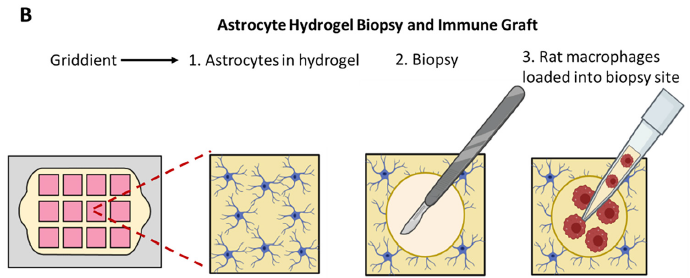
Figure 4: Biopsy and cell grafting experiment. (Image © 2023 The Royal Society of Chemistry)

The importance of these results is significant. The research team has shown that it can monitor the health and location of astrocyte cells with a number of analytical techniques including live- and dead-cell fluorescent tagging, and monitoring of gene regulation. It has also demonstrated that their attempt at tissue grafting showed successful cell penetration. This at least means that their brain-on-a-chip device should be tested more rigorously to explore its capabilities on several fronts — for example, monitoring movement and viability of different cells within a variety of tissues/proxies, evaluating the effectiveness of emerging, unproven stroke therapeutic treatments, etc. Furthering this research could provide invaluable insight into stroke tissue damage and regeneration techniques. Also, there is no reason to think that a similar approach could not be taken with other tissue models to characterise diseases and conditions, and evaluate therapies.

Survey: metrology in microfluidics

Survey: metrology in microfluidics

The Microfluidics Association is participating in a workshop on November 13th-14th, 2023 focussed on on metrology in microfluidics, and hosted by the metrology wing of the French Cetiat institution. In preparation, we at the Microfluidics Association (I’m on the Board) want to brush up on what the microfluidics community wants and needs as regards metrology. Here’s a letter from Henne van Heeren, fellow board member.


“For our work in developing standards and metrology protocols it is crucial that we know what the community needs. Therefor you you are invited to participate in our survey concerning prioritizing metrology and standardization work for the coming years.
Your participation in this study is completely voluntary, so if you feel uncomfortable answering any questions, you can skip questions or withdraw from the survey at any point.
The information you will supply will be coded and will remain strictly confidential.

Thank you very much for your time and support. Please start with the survey now by clicking on https://www.surveymonkey.com/r/D6RF9JM

This survey is held in preparation for the November workshop “on the road to standardization in microfluidics and organ-on-chip”. For more information on this workshop, please click here.

Best regards on behalf of the Microfluidics Association

Henne van Heeren
henne@enablingMNT.com
0031 78 6300748

Download our published whitepapers: https://microfluidics-association.org

We acknowledge the support of the MFMET project, https://mfmet.eu/ and the European Metrology Programme for Innovation and Research: EMPIR.”


On behalf of the Microfluidics Association, I would like to encourage any and all working in microfluidics to provide your input about metrology via our survey. I gave my 2¢ this morning! 🙂

Sensitive microfluidic detection of dopamine

Sensitive microfluidic detection of dopamine

Impressive detection of very low concentrations of the neurotransmitter dopamine have just been demonstrated in research coming from the Wang and Tu research groups at the Department of Bioscience and Biotechnology at the National Taiwan Ocean University and the Institute of Chemistry at the Academia Sinica, respectively, in Taiwan. The work was recently published as an on-line article in ACS Sensors.

Figure 1: chemical structure of 3,4-dihydroxyphenethylamine, or dopamine (Source: Wikipedia)

Dopamine, shown at right for like-minded chemistry nerds, is a small molecule that acts as a neurotransmitter in the brain and as a hormone with different functions elsewhere in the body including blood vessels, kidneys, the pancreas and the immune system. The ability to detect dopamine is of great interest, since abnormal dopamine levels are indicative of neurological conditions such as depression, schizophrenia, attention deficit hyperactivity disorder (ADHD) and Parkinson’s disease.

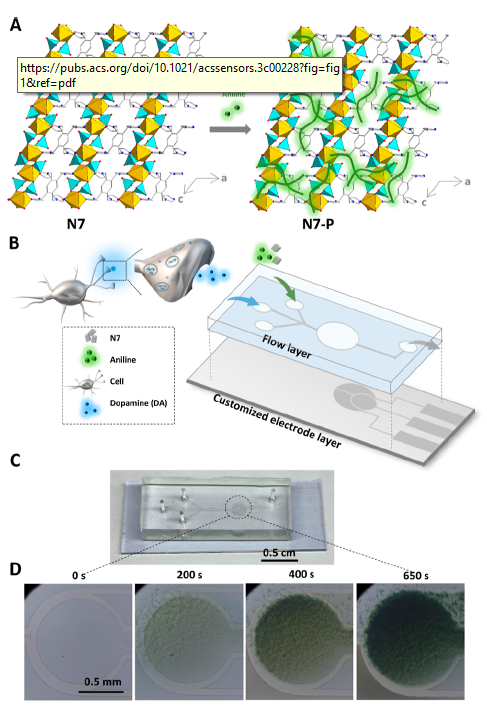
Figure 2: coating material, device structure and electropolymerisation of working electrode (Image © 2023 American Chemical Society)

The researchers created a microfluidic device to act as the dopamine biosensor. The glass base layer had an indium tin oxide (ITO) coating patterned with a CO2 laser to form the 3-electrodes used for electrochemical detection. The fluidic cover was made by soft lithography: cast moulding of PDMS over a photolithographically patterned SU-8 master. An additional novel coating was applied to the working electrode to afford the selective sensing of dopamine. The coating, “N7-P”, was electro-polymerised via cyclic voltammetry from a solution of an indium phosphate-organic linker hybrid and analine. The graphic at right from their paper shows a) the structure of the yellow/cyan InPO4/organic linker crystalline structure (left) with added green polyaniline (right), b) electrode base (grey) and fluidic cover (blue), together with N7-P coating materials input well (top) and neuroblastoma cell/dopamine input well (middle), c) photo of assembled chip, and d) microscope imagery of working electrode during coating process of the N7-P.

Performance demonstrated by the groups’ microfluidic dopamine sensors was impressive in terms of the low detection limits and dynamic ranges seen in static and flowing configurations. With no flow, the linear range was 1 pM to 10 nM, while under flow (optimised at 50 µL/h), the range was 1 aM to 100 fM, with an LoD of 0.183 aM. Unfortunately, the authors did not disclose the dimensions of the device’s fluidic channels and detection chamber, which become significant when low concentration levels approach the need for single molecule detection, as in this case.

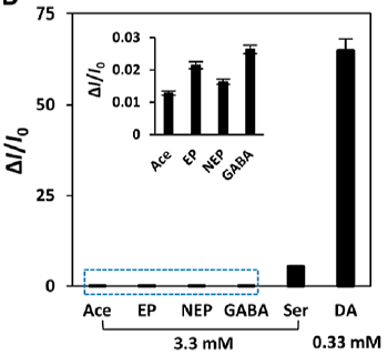
Figure 3: selectivity for dopamine vs. other interfering neurotransmitters (Image © 2023 American Chemical Society)

Also important was the excellent selectivity shown by the devices for sensing dopamine in the presence of five other neurotransmitters with similar structures — acetylcholine, epinephrine, norepinephrine, gamma-aminotbutyric acid and serotonin. A plot showing the amperometric response of the sensor to dopamine (DA) versus these other five potential interferents at 10-fold concentrations (labelled Ace, EP, NEP, GABA and Ser, respectively) is shown at right.

In their experiments looking at biologically produced dopamine from rat cells (> 1000 cells/mL), the concentrations measured by the microfluidic biosensor tracked very closely with the results obtained using a commercial ELISA system when larger numbers of cells were used (> 1000 cells/mL); no comparison of results could be made for smaller numbers, as the commercial system failed to generate any signal. Signal from as few as 21 cells could be sensed at dopamine concentrations of ~15 aM.

If I’ve lost you with a bunch of chemistry concentrations jargon, please accept my apologies! The salient point is, the system has exquisite sensitivity, and that can be very useful in many situations where e.g. human biopsy or research cell samples are very small, and the impact of the cells’ environment or the effectiveness of a therapeutic drug for a neurological disorder is being evaluated. Having more sensitivity is like having more horsepower: it’s (almost) always a good thing!

Another important point is that the devices are manufacturable with what look to be fairly mainstream or easily implemented microfabrication methods. They mention that the closest rival electrode coating material to afford the selectivity for dopamine are “metal organic frameworks”, or MOFs. In a table they provide (Table S2 of their supplementary information), the current biosensor’s linear range is at least 8 orders of magnitude (i.e. 80 million times) more sensitive than that seen with MOF-based electrochemical detection. It’s not clear how ease-of-microfabrication compares between MOFs and the present coating technology.

Microfabrication of Zn-air microbatteries via 3-D printing

Microfabrication of Zn-air microbatteries via 3-D printing
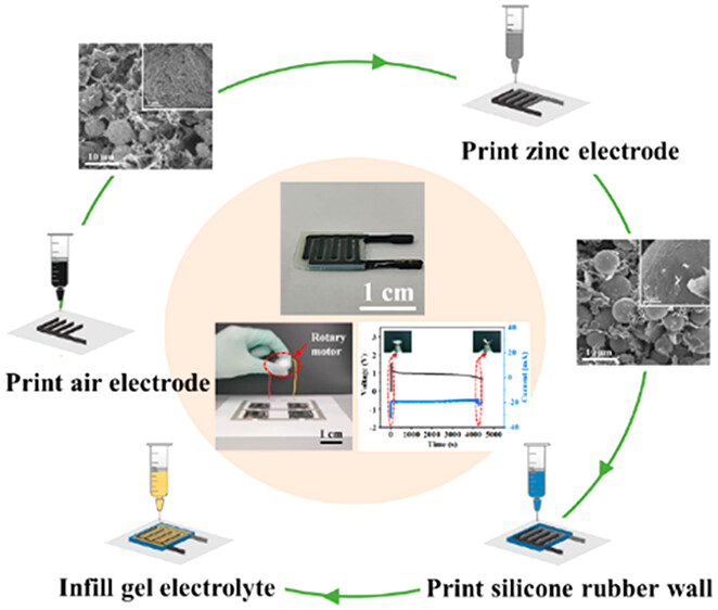
Figure 1: 3-D printing of 2 interdigitated electrodes, silicone enclosure and gel electrolyte components of battery (Image © 2023 American Chemical Society)

Guoxian Li and Chuizhou Meng’s groups in Mechanical Engineering at the Hebei University of Technology recently published an interesting article pre-print from the American Chemical Society’s Applied Materials & Interfaces. Their research paper shows the simple microfabrication of rechargeable batteries in the form of 3-D printed interdigitated electrodes (IDEs) on a variety of substrates. The appearance and simplified fabrication process of the batteries are illustrated in Figure 1 above right.

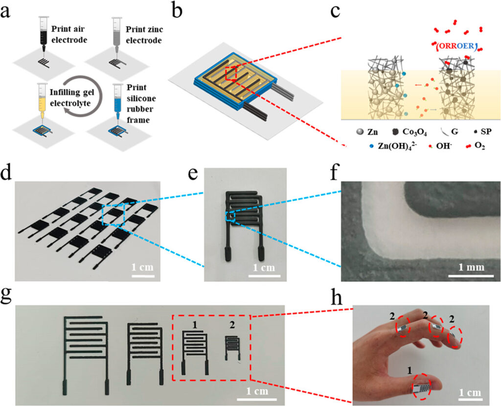
The paper and supplementary information provide a fair amount of detail regarding the fabrication process used to make the electrodes. Four slurries were prepared for the air electrode, zinc electrode, interelectrode gel polymer electrolyte, and silicone rubber to close the cell. Figure 2 below left shows the printing process, chemical composition, sizes and examples of the IDE batteries. The air electrode incorporated tricobalt tetraoxide (Co3O4) to catalyse the oxygen reduction during discharge and oxidation (evolution) during charging.

Figure 2: Microfabrication via 3-D printing (a), electrode layout & composition (b-c) as well as photographs of IDE batteries of different sizes (d-h). (Image © 2023 American Chemical Society)

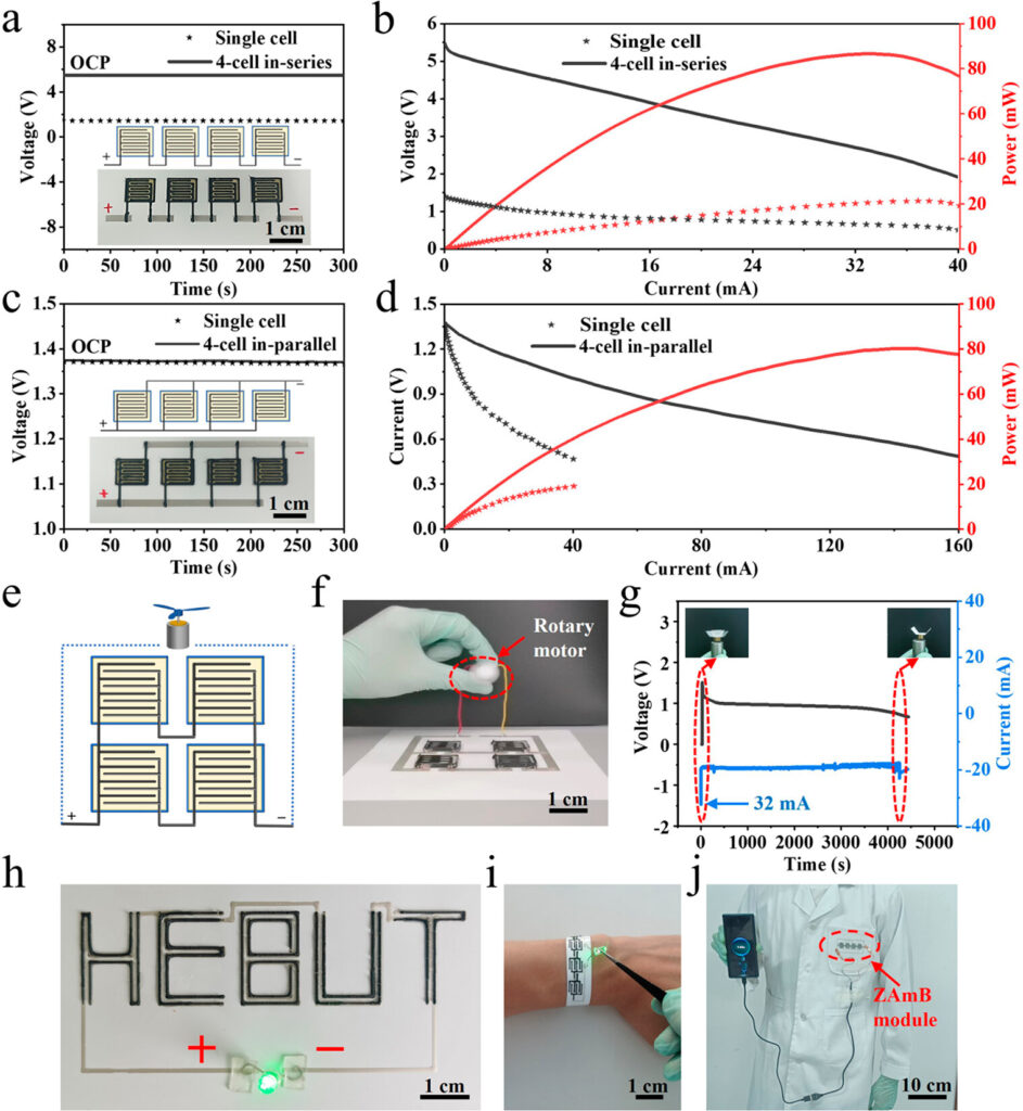
Different sizes (x, y) and thicknesses (z, depending on the number of layers deposited) of IDEs were prepared and characterised; electrode arrays were on the order of 1 cm x 1 cm, with thicknesses on the order of 1.5 mm. Arrangements with batteries in parallel (high current and power), in series (high voltage) or both were evaluated. Figure 3 below shows four batteries arranged in series (a), in parallel (c), and the polarisation & power curves for the single and 4-battery arrangements in each case for series and parallel circuits (b & d, respectively). Open circuit potential (i.e. voltage with no load) of the battery arrangements are also shown in (a) and (c). They claim high power density performance at 77.2 mW•h/cm². Lifetime of full power output varied approximately with the number of layers deposited.

Several examples of printing options and applications were also provided, demonstrating the use of the batteries in powering a fan motor (Figure 3, e-g), LEDs (h-i) and charging a cell phone (j). Interestingly, different Zn/air electrode alignments are possible, as in (h), as are different substrates. Normally, polyethylene terephthalate (PET, a common thermoplastic used for blowmoulding e.g. pop bottles, clamshell produce packages, etc.) in a flat sheet is used, but flexible wristbands (i) and cloth patches (j) were also demonstrated.

Figure 3: Serial and parallel battery circuits and performance (a to d), and application examples to power fans, LEDs and recharge cell phones (e-j). (Image © 2023 American Chemical Society)

This technology shows significant promise. The use of cobalt oxide as a catalyst and zinc as a metal are important advantages. Compared to the use of expensive noble metals like platinum and ruthenium, the use of Co3O4 as a catalyst is both more economical and environmentally sustainable. Likewise, the authors and others point out that zinc-air batteries have about five times the energy density of lithium-ion batteries, and are a potentially greener alternative. I was also struck by several potential benefits in the context of MEMS and microfluidic devices. The ability to integrate microfabricated batteries to power on-board components, without requiring an additional button cell, presents more opportunities. The battery size (capacity) can be tailored to device needs, reducing costs and waste impact for single-use scenarios. Also, the fact that these batteries are rechargeable opens the door to the development of multi-use or longer use MEMS and microfluidic devices, which can significantly decrease COGS and environmental footprint in an eventual product.

Reproducible pH control in microfluidic chambers for combinatorial chemistry

Reproducible pH control in microfluidic chambers for combinatorial chemistry

Researchers from César Pascual García’s group at the Luxembourg Institute of Science and Technology as well as Wouter Olthuis from the MESA+ Institute at the University of Twente in the Netherlands have designed, built and characterised a microfluidic device capable of controlling pH between acidic and neutral pH values. Their work was recently published on-line as an ACS Omega pre-print article.

Why does on-chip pH control matter? Control of acidity allows many molecular states and reactions to be regulated, e.g. for the synthesis of oligonucleotides (DNA), polypeptides (proteins), saccharides (carbohydrates), all of which are key in many areas of analytical, organic and pharmaceutical chemistry. One important area is combinatorial chemistry, which allows an array of related chemical compounds to be meticulously synthesised, and then evaluated against a target molecule for a given purpose, such as therapeutic effect of a drug, or receptor affinity as a bio marker of a disease or condition to be diagnosed. Regulating a large number of such reactions in a dense matrix array aboard a microfluidic chip means that many more reaction options can be screened simultaneously with very small amounts of reagents in an automated fashion, making for more effective drugs and diagnostics with lower development costs.

Their microfluidic devices have a Si/SiO2 base with Pt working, counter and reference electrodes, SU-8 patterned fluidic channels and removable glass lid. Ti/Au electrodes were lithographically patterned using lift-off, with subsequent electrochemical deposition of Pt and functionalisation with the electro-active species 4-aminothiophenol (4-ATP) thereafter. A photo of a 4-cell arrangement of their system, pH response and reversible reaction controlling the chamber pH are shown below.

Left: two of the four three-electrode cells used to adjust pH. Middle: trace depicting the alternating bias applied to the two cells, and resulting pH response. Right: electroactive species 4-ATP shown reacting reversibly between oxidised (purple) and reduced (red) configurations. (Image © American Chemical Society/Divya Balakrishnan et al.)

Fairly reproducible control of pH through 100+ cycles was achieved, with pH stability lasting through holding times of up to 10 minutes. pH can also be tailored to given values between pH = 3-7 ±0.4, based on the fluorescence intensity of the carboxy semi-napthorhodafluors dye used to validate performance.

The authors have taken previous work by others using photoactive compounds for pH regulation ahead a step, by removing the need for optical interrogation of the cell. This eliminates expensive optics instrumentation and alignment issues, replacing them with mass-manufacturable electrochemical control. While the current design is not configured for high density, the 2.5 nL reaction volumes are already quite small, and it is not hard to imagine a high density layout for manufacturing. Application of this technology to the development of therapeutic drug candidates, disease diagnostics and other areas seems promising.

Zeon acquires Edge Precision Manufacturing

Zeon acquires Edge Precision Manufacturing

According to recent LinkedIn posts and a press release, polymer manufacturing leader Zeon Specialty Materials Inc. (subsidiary of Zeon Corporation, Japan) recently acquired Edge Precision Manufacturing, a microfabrication manufacturing house based in the Boston, MA area. In the world of microfluidics, Zeon is a well known manufacturer of its cyclic olefin copolymers (COPs) Zeonor® and Zeonex®, widely used in the injection moulding of microfluidic products.

The manufacture of plastic microfluidic devices is most often performed with injection moulding, given its economic edge at high volumes, and less often with hot embossing, given its slower throughput, however hot embossing has the edge for devices with more demanding fine features and fidelity requirements. Edge Precision Manufacturing, formerly Edge Embossing, is able to combine the best of both worlds with a high volume, high precision hybrid approach, where hot embossing is used for the most demanding areas of a component, and injection moulding for the more routine feature sizes.

Zeon’s acquisition of Edge Precision Manufacturing is the latest in a series of microfluidics and MEMS fab house acquisitions that has seen Invenios acquired by Corning and thinXXS acquired by IDEX in 2017, miniFAB acquired by Schott and Micralyne acquired by Teledyne in 2019, and most recently microLIQUID acquired by TE Connectivity in 2021.