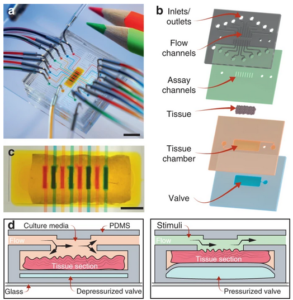
 Interesting research regarding a microfluidic chip that facilitates multiplex analysis on a single biopsy tissue sample was recently published in a Microsystems & Nanoengineering article from José Luis García-Cordero’s group at Unidad Monterrey. The technique allows highly irregular surfaces to be simply examined without further modification.
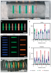
The PDMS chip, shown in upper image (©Nature – Microsystems & Nanoengineering), uses a ~3 x 7 mm chamber for the biopsy tissue sample, allows media to perfuse around the sample (orange fluid). Once the valve is pneumatically actuated (7-35 kPa), the tissue is pressed against the lid to seal the microfluidic channels and allow the simultaneous introduction of discrete drug or tissue culture solutions through each of the eight channels. Characterisation studies shown in the lower image (©Nature – Microsystems & Nanoengineering) demonstrate good permanent sealing and no cross-talk between channels. Also, the continuous periperfusion of media around the sample increased tissue cell viability over the first several hours in the microfluidic device vs. in a petri dish.
The authors discuss the possibility of using biopsy samples in devices to perform drug efficacy, safety and toxicology studies before clinical trials, as well as facilitating research in physiology, metabolism and tissue regeneration.
