Open channel microfluidics

Open channel microfluidics

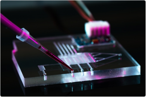
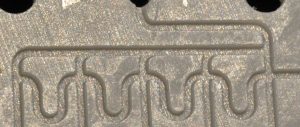
theberge-open-chnl-uf-1An interesting article from Ashleigh Théberge’s group at the University of Washington reviews the relatively recent introduction of open capillary microfluidic systems (as distinct from electrowetted digital microfluidics).  The paper was recently pre-published in Analytical Chemistry as an ASAP article.
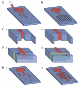
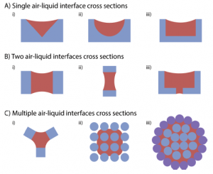
theberge-open-chnl-uf-2The authors review a number of different channel geometries and fibre bundle configurations that can be used to promote open capillary flow.  The helpfully provide the equations that use channel geometry and contact angle to determine whether capillary flow is energetically favourable.  This is done for both conventional monolithic (single material) channels, as well as composite material channels.

Pros and cons of the open capillary approach are surveyed.  The authors list several advantages:

  1. simplified fabrication by obviating the need for bonding and potential associated use of solvents on the substrate, process development/trade secrets, manufacturing cost, etc.;
  2. ease of performing surface modifications, such as for hydrophilicity/hydrophobicity, silanisation or other derivitisation, blanket or patterned exposure to UV, plasmas, chemical or physical vapour depositions (PVD & CVD), application of delicate bio-reagents that can’t withstand thermal bonding;
  3. accessibility of channels for adding or removing reagents or components with pipettes, tweezers (as for tissue scaffolds)
  4. elimination of air bubble issues, due to the open interface.

They also mention disadvantages primarily stemming from the open channel access such as higher evaporation, evolution and/or exchange of dissolved gases, liquid leaks to non-channel paths, and the inability to generate higher pressures in channels (beyond those of capillarity) and thus use valves, etc.

Lastly, a number of different applications are noted, though all appear to be academic in nature at this stage.  While I see the advantages of flexibility and reduced manufacturing cost offered by the open channel concept, I wonder how a product would be able to mitigate against evaporation and contamination issues in viable approach suitable for a robust consumable suitable for untrained users.  Perhaps the authors have the answers; they mention that they have financial interests in two companies, Salus Discovery and Stacks to the Future, involved in the commercialisation and IP related to some of the technologies presented.

Dendrite cell chemotaxis in microfluidic mazes

Dendrite cell chemotaxis in microfluidic mazes

korean-chemotaxis

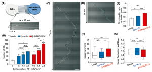
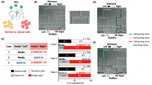
South Korean researchers have recently shown that immature dendrite cells undergo chemotactic migration through microfluidic mazes preferentially towards healthy or cancerous cells versus cell-free medium.  The new research findings, published in a Lab on a Chip article, come from Cho’s group at the Institute for Basic Science, Grybowski’s group at the Ulsan National Institute of Science and Technology, and Jeon’s group at the Pohang University of Science and Technology.

Chemotaxis is the movement of cells towards or away from chemical stimulus (attractants or repellents, respectively).  Bacteria accomplish this through biased ‘random’ walk cycles, where the cells use their flagella to move in a given direction, then stop and sense whether they have moved up or down the stimulant’s concentration gradient to determine subsequent reorientation and straight-line translation.  Migration towards attractants by dendrite cells (surveillance agents and messengers for the immune system) is well documented for mature but not immature dendrite cells.

In this study, immature cells were allowed to migrate towards cell medium (control), EpH4-Ev healthy cells or beta-MEKDD 116 cancer cells.  In one experiment series, comparisons in migration were evaluated by allowing the immature dendrite cells to migrate from a single inlet towards either of two outlets that contained two of the three cell attractants.  Attraction bias was clearly shown to be (beta-MEKDD 116) > (EpH4-Ev) > (cell medium).  In another series of experiments, different cytokines drawn from the cancerous beta-MEKDD 116 cells were compared, and the protein Gas6 was found to have the largest attractive effect.  Large numbers of replicate analyses allowed the authors to nicely quantify the confidence limits that applied to their results.korean-chemotaxis2


First image of black hole: epitome of scientific collaboration

First image of black hole: epitome of scientific collaboration

black-hole-different-observatories Accomplishements in microfluidics and analytical chemistry are normally front and centre in this blog, but this is a nod to some great work in physics.  It turns out physics is more complicated than F = ma. 😉

There was a big splash in the media around April 10th about the first images of a black hole ever obtained, and they are indeed pretty fascinating.  What is at least as fascinating is the coordinated, collaborative effort between the astrophysics teams in several countries to generate these images.  A few editorial pieces summarise the tremendous scope of the work nicely on webpages at MIT, the Event Horizon Telescope (or EHT), and publisher IOP.  There are six open-access publications listed at the bottom of the EHT page that describe the work and its results.

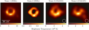
The teams all belong to the EHT which is an array of radiofrequency telescopes that work in harmony to image a given target at the same time.  The resolution afforded by teaming the individual telescopes together is vastly better than that of any individual telescope.  A few images from papers that were published simultaneously on April 10 show some of the results.  The first shows several pictures of the M87 black hole taken from different observatories early in the project, before many of the efforts at noise reduction were implemented (image from Figure 4 of this paper).  array-of-different-observatoriesThe second shows the location of the different observatories in Europe, North and South America, Hawaii and Antarctica that were teamed together for the effort (image from Figure 1 of this paper).

To be able to work synchronously, all the observatories had to use precise timestamping of their images with atomic clocks.  Each observatory generated so much data, about 1 PB (PB = petabyte = 1 million GB), that it was faster to simply fly the hard drives to the Max Planck Institute for Radio Astronomy (Germany) and MIT’s Haystack Observatory (Boston, US) for the data processing.  Interestingly, the images black-hole-different-dayschange appreciably from day to day, as shown below (image from figure 15 of this paper).
Any collaborative scientific effort of this size is remarkable, all the more so given the size of the groups in different countries, funding sources from yet many more countries, etc.


Microfluidic microneedle PoC system created by laser machining

Microfluidic microneedle PoC system created by laser machining

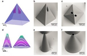
german-microneedlesA recent article in Nature: Microsystems & Nanoengineering by Ralf Hellmann’s group at the Aschaffenburg University of Applied Sciences and Thomas Walther’s group at the Technische Universität Darmstadt in Germany outlines impressive laser machined microfluidic microneedle devices.

Devices were fabricated in PMMA coated in OrmoComp® photoresist in a two-step process.  Direct laser written photoresist microneedle arrays (tip radius of 13-21 µm) and 3-D microfluidic PMMA channels were combined in a device using a single femtosecond pulsed laser for fabrication.  The fabrication process is claimed to be simpler than traditional fabrication methods, but unfortunately, no throughput figures were provided.  It would be nice to know what these are to understand how readily this fabrication technique could be applied as a manufacturing tool.

Devices were characterised with force vs. time curves for injections of a rhodamine B test solution through pork skin (which shares similar morphology to human skin) and to verify microneedle integrity during use.

The authors highlight that the devices could provide a significant leap forward for point-of-care applications including drug delivery and diagnostics.

Microfluidic screening of bacteria for power generation, waste digestion

Microfluidic screening of bacteria for power generation, waste digestion

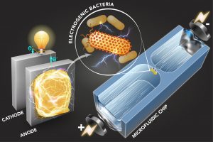
mit-electric-bacteria-pressNew research out of Cullen Buie‘s lab at MIT’s LEMI lab shows effective sorting of different strains of bacteria using microfluidics-based dielectrophoretic (DEP) analysis.  The research was published in Science Advances article (DOI: 10.1126/sciadv.aat5664) and also profiled by MIT News; a second MIT news article provides some background to microbial digestion and power generation.

The cell manipulation works by virtue of a DEP trap. A cell suspension flows through a small orifice (~ 50 µm wide) between two microfluidic chambers under an electric field.  The electroosmotic and electrophoretic forces generated by the field determine each cell’s speed in the bulk solution.  At the constriction, there is additionally a dielectrophoretic force that acts to trap cells of a certain polarisability, depending on the applied field strength.  Since polarisability relates to extracellular electron transfer (EET), or a cell’s tendency to generate electricity during respiration, the trap can act as a filtre or screen for bacterial strains based on their tendency to generate electricity.  In other words, different strains can be selectively trapped or screened based on the applied voltage.

The value of being able to discern between the different strains of bacterial in terms of their tendency to generate power while digesting the components in wastewater can hardly be understated.  Apparently 3 of energy in the US is spent on wastewater treatment, while the wastewater itself contains 10 times the energy required for its own processing.

ISS microfluidic experiments to reveal secrets of aging

ISS microfluidic experiments to reveal secrets of aging

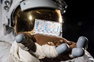
iss-nasa-organ-on-a-chipA Technology Networks article based on NIH-funded research describes how organ-on-a-chip microfluidic devices just reached the US’ NASA portion of the International Space Station (ISS), the ISS National Lab, last Wednesday as part of an investigation into the effects of aging, mimicked by weightlessness, on the immune system.  The research team is led by Professor Sonja Schrepfer at UC-SF.

The immune system chips incorporate immune, bone marrow and blood vessel cells.  The physiological effects of space flight on astronauts, and the tissues on these chips, are similar to those of aging, including bone and muscle loss and altered immune systems.  The several dozen chips will be incubated for 2 weeks, and then frozen until they return to Earth for analysis at the Schrepfer lab.  Future SpaceX re-supply missions to the ISS in March and April 2019 will bring kidney, bone/cartilage, and blood/brain barrier chip sets as part of the same research programme.

It is hoped that the research may provide better understanding of, and potentially improved therapies for the process of aging.

Alzheimer’s disease modelling using brain-on-a-chip

Alzheimer’s disease modelling using brain-on-a-chip

alzheimers-on-a-chipImpressive research relating to Alzheimer’s disease (AD) research on a microfluidic platform recently emerged from the Cho group at the University of North Carolina at Charlotte and the Tanzi group at the Massachusetts General Hospital & Harvard Medical School; it was published in Nature Neuroscience and highlighted in a News Medical Lifesciences article.

The work describes a flexible microfluidic system that allows the effectiveness of different AD treatment drug compounds to be evaluated based on the responses of different types of brain cells, particularly the microglia cells involved in the brain & CNS immune response.  The microfluidic device contains 2 chambers, one with neuron cells and astrocytes, and the other with microglia.  Reduced microglia ‘recruitment’ by the other cells, as visualised in the channels between the chambers, via excreted cytokines by the neuron and astrocyte cells indicates reduced neuronal damage and, presumably, reduced loss of memory or other mental capacity.

There are over 3000 approved drug compounds for treatment of Alzheimer’s disease, according to Joseph Park, first author on the research paper, so evaluation in a representative biological and disease environment such as created in their chip is crucial.  Human trials are lengthy, expensive and carry risk for the participants.  Selection of the most promising candidates for drug therapy through understanding their interaction with brain cells in this microfluidic system offers promise to accelerate the pace towards effective treatments of Alzheimer’s disease.

BioFluidica’s microfluidic tumour detection technology

BioFluidica’s microfluidic tumour detection technology

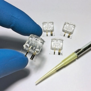
biofluidica-deviceA recent article by Bradley Fikes in the San Diego Union-Tribune highlighted the progress achieved by a local leading biotech company, BioFluidica.  Founded by Professor Steve Soper at Kansas University and headed up by CEO Dr. Rolf Muller, two veterans in the area of microfluidics research and product development, the company is effectively translating the inherent capabilities of lab-on-a-chip technology into the foundation of their cancer-screening products.

BioFluidica uses injection moulding to economically mass produce their plastic microfluidics chips.  While injection moulding has been effectively applied to microfluidic products with ‘normal’ channel geometries (width similar to depth), BioFluidica’s device channels appear to be tall and thin, making high fidelity, high volume reproduction challenging.  That they can use injection moulding for these devices is no mean feat; it is not a surprise to hear that they had to go through several manufacturers before finding one in Austria equal to the task

The devices are used to perform affinity-based assays of circulating tumour cells (CTCs), and work directly with whole blood, a distinct advantage in terms of cost and ease-of use, and also likely in terms of the quality of the assay results.  BioFluidica estimates that lung biopsy screens would be reduced from $10,000 USD to roughly $5,000 using their technology.

Simple but elegant microfluidic cell concentrator

Simple but elegant microfluidic cell concentrator

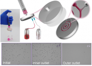
cell-concentratorResearch from Zhonghua Ni‘s group at Southeast University in China published recently in Analytical Chemistry (DOI: 10.1021/acs.analchem.8b02201) allows for effective cell concentration along a spiral microfluidic channel.

The device is connected directly to a syringe, and outward flow in the microfluidic spiral channel is generated simply by the operator pushing on the plunger.  Separation by inertial (centrifugal) forces partitions the cells to the inside channel ‘track’ such that a Y-shaped bifurcation at the outer spiral terminus yields a concentrated cell suspension on the inner outlet and almost cell-free solution on the outer outlet.

The device embodies simplicity as a virtue.  It has no active components, and requires no electrical power, only average human thumb power :-); it simply requires a sample-filled syringe and two collection vials.  It is well conceived for both resource-poor and lab environments alike.  Their bold claim that one could bring their concentrator “into commercial outcomes without additional redesigning” initially made me chuckle, but after absorbing their work, I have to admit they appear to be exaggerating only slightly!  An Abbott, BD or start-up would require some industrial product design, but technically, they appear to be at the finish line.  I can’t speak to the competition in this product segment, and sure hope they have a good patent, but it seems pretty clever!

Flexible & economical lab-on-a-chip platforms

Flexible & economical lab-on-a-chip platforms

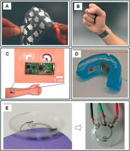
flexible-loac-1An interesting review of lab-on-a-chip devices made on flexible substrates was recently published in Lab on a Chip by Anastasios Economou and Christos Kokkinos at the University of Athens, and Mamas Prodromidis at the University of Ioannina.  The authors cover devices made on polymer, paper and textile fabric substrates and which use electrochemical biosensing for detection.  Lab-on-a-chip & microfluidic devices based on flexible substrates are very attractive given the low costs of the substrate and deposited materials, inexpensive and well established high volume manufacturing processes, and physical flexibility as an attribute for body-wearable devices.

Sensor/device formats include patches, bandages and wristbands for dermal/transdermal sensing, dental mouthgards for salivary analyses, tooth patches for bacterial detection and contact lenses for tear fluid analysis.  Analytical applications include immunosensing, glucose monitoring, bovine serum albumin (BSA) sensing, metabolite sensing, cancer marker detection and HIV detection.  Deposition methods include lower-cost approaches such as screen printing, inkjet printing and electrodeposition for materials such as graphite, indium tin oxide, polyethylene terephthalate (PET).  Polymer substrates include thermoplastics like polyethylene, polystyrene, PET, polypropylene, polycarbonate, cyclic olefin (co)polymers (COC & COP), thermoset polymers like polyimide, and photoresists.  Filtre paper is the most common type of paper substrate, though newsprint is also mentioned.  3-D, folding or origami approaches are also discussed, as previously highlighted in this blog.

HJC Consulting Product Profile Award

HJC Consulting Product Profile Award

HJC Consulting is pleased to launch a newd90_dsc_0575-cu monthly Product Profile Award series to help give microfluidic product developers a leg up in creating awareness about current or upcoming products. The award will provide that month’s successful applicant a chance to get the word out through a product on HJC Consulting’s website and social media channels regarding their microfluidic or lab-on-a-chip product’s milestones such as a launch, revision with enhanced performance, new product research or patent publication, etc.

The profile will be awarded, free of charge, to one submission per month.  To be eligible, a profile submission must:

  • speak to the technical method of operation and capabilities of the product;
  • include imagery (photos, data, logo, etc.) for which copyright is owned;
  • be free of copyrighted, confidential or proprietary information;
  • highlight preferred applications and/or targeted market segments;
  • include details concerning:
    • (co-) developer & (co-) author email addresses, and social media addresses if desired;
    • company physical address, contact & phone number, website & media release URLs, social media addresses for Facebook, Twitter and LinkedIn; and
  • be 300 words or less.

See examples of current blog posts for guidance.

The profile will be hosted on the HJC Consulting blog, and will be posted simultaneously through HJC Consulting’s social media accounts.  Suggestions for social media post content is also welcomed.

Please send queries or submissions via email to John Crabtree.  Successful submissions may be edited for content and length by HJC Consulting.  Unsuccessful submissions may be considered for subsequent months, if desired.

We hope to see your application soon!

Sepsis detection via microfluidic neutrophil analysis from Massachusetts General Hospital & Harvard Medical School

Sepsis detection via microfluidic neutrophil analysis from Massachusetts General Hospital & Harvard Medical School

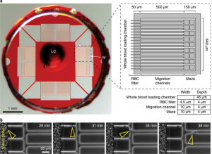
mgh-neutrophil-analyser-for-sepsisFascinating new research by Felix Ellett and co-authors from Massachusetts General Hospital (MGH) and Harvard Medical School (HMS) shows elegant detection of sepsis in hospital patients via microfluidic blood analysis.  The study was published recently in Nature – Biomedical Engineering and profiled by MGH and Technology Networks.

Previous research from Daniel Irimia‘s group at MGH and HMS in 2014 showed that dysfunctional behaviour of neutrophils (a type of white blood cell) correlated well with sepsis.  This current research from his and Jarone Lee‘s (MGH) groups takes the next step and uses a microfluidic device to perform the analysis.  Their 5-mm device has 4.5-µm channel filtres (to screen out red blood cells) and meandering channel networks to allow the neutrophils to leave the sample drop of blood, and be observed in the microfluidic channel network.  Discrimination by neutrophil characteristics & behaviours (such as number of neutrophils, oscillations and motionless time in microfluidic channels, migration distance and tendency to migrate back to the central sample chamber) allowed the researchers to correctly predict the presence of sepsis in over 95% of patients in a small study of 19 ICU patients.

Detection of sepsis in patients is of critical importance.  According to the MGH and TN article profiles, about one million cases of sepsis occur annually in the US, with 25% of cases being fatal.  Comparable data from the UK showed that the mortality rate for hospital admissions with severe sepsis assessed withing the first 24 hours was 45%.  Current detection methods leave much to be desired, with misdiagnosis occurring in 30% of patients.  The arrival of fast, accurate diagnostic methods would lead to better patient outcomes, likely including saved lives.

Bullish microfluidics market projections by MarketsandMarkets

Bullish microfluidics market projections by MarketsandMarkets

A recent report by Markets and Markets cited in an article by The Health Star shows aggressive microfluidics product market growth predictions that exceed previous strong predictions from other research enterprises.

Markets and Markets put the 2017 market value at $8.28 B USD with a compound annual growth rate (CAGR) of 22.6% forecasted until 2023.  Other research firms, however, have lower estimates of both the current market as well growth predictions for similar time spans.  Yole Dévelopment’s 2017 report, for example, estimated the 2016 market to be ~$2.1 B USD, and projected a CAGR of 18% out to 2022.  A 2017 report by Transparency Market Research put the 2016 market at $4.76 B USD and CAGR at 11.4% through 2025.  Future Market Insights’ 2017 report on the point-of-care (PoC) segment of the microfluidics market, the largest segment in their view, estimates this segment to have been $1.0 B USD in 2016, and to grow at a 14.5% CAGR until 2026.

The reason for the large differences between Markets and Markets’ estimates and those of others – roughly, 2-4x larger market values and 1.25-2x larger CAGRs – is not clear, but may be due to definition of the market scope, i.e. what products are considered “microfluidic”.

Great Customer Service from Christie-Phoenix & CFC Underwriting

Great Customer Service from Christie-Phoenix & CFC Underwriting

I don’t hesitate to bring poor customer service into sharp focus with a service provider, and with the wider world, if necessary … so it seems that I should also equally underscore excellent service!  I had just such an experience recently with my former commercial insurance broker, Christie-Phoenix, part of Arthur J. Gallagher Canada, and former underwriter, CFC Underwriting.

It started with me leaving them because I’d been able to secure a better deal through a professional affiliation.  My broker with Christie-Phoenix, Ted Stelck, was very helpful all the way through the process, first getting me competitive quotes, and then once I’d decided to leave, ensuring I was properly covered during the transition.  I wound up changing firms only 20 days into my new annual policy, and eventually an invoice trickled in for coverage during this period.  To my surprise, it included a minimum charge of 1/3 of the annual premium, instead of a pro-rated charge based on time covered, as is the case with e.g. home or auto insurance.

I raised my objections regarding this heavy minimum ‘administrative’ levy with Ted, who passed it along to his CFC policy counterparts, who confirmed that this was how the charges had to be.  I got him to ask again to see if they would make an exception, but to no avail.  The CFC policy people pointed out that the fine print on page thirty-something allowed for this.  Annoyed, I finally woke up and contacted the CFC customer relations people through Facebook, where I described my situation and displeasure.  I had an email and phone call within 24h from a Gillian Harvey requesting that I kindly provide her with a full description of the situation.  Gillian listened attentively, and made it clear that, while the policy allowed for the ~1/3 premium fee, she totally understood my annoyance as a customer, and pledged to do something about it.  A day later, I had a letter wiping the whole fee, and also covering the broker’s fee, as a gesture of goodwill.

Nicely done, Christie-Phoenix and CFC Underwriting – way to make it right!!  I’ll now consider this situation as an example of a good outcome when the rubber hit the road.  I’m also publicising this so that my circle of contacts can appreciate that you provided good customer service even when you’d lost the business.  In my books, this is a great example of putting the customer first, and like most people, I strive to give my business, and kudos, to companies that can demonstrate their understanding of this concept.

Single bacteria cell microfluidic analyser

Single bacteria cell microfluidic analyser

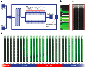
ubasel-bacteria-loac-deviceGround-breaking lab-on-a-chip research out of the van Nimwegen lab at the University of Basel, Switzerland and the Myers lab at the Max Planck Institute of Molecular Cell Biology and Genetics, Germany, has enabled both rapid environmental control of cell media, and the ability simultaneously monitor large numbers of bacteria as single cells.  The work, recently published in Nature Communications and highlighted in Technology Networks, describes a PDMS microfluidic device with ~1900 channels measuring ~ 1 x 1 x 25 µm that house the single cells, and which are connected perpendicularly to larger supply channels containing the cell medium solution.  The device also features continuously variable 2-input control of cell media via a split channel configuration, together with a mixing serpentine upstream of the smaller single cell channels.  New image analysis software was also created to handle the enormous amount of image data generated by this system when in operation.

The device is intended to provide a real-time monitoring window into gene regulation in bacteria, a laudable advance.  For example, bacterial response to different antibiotics can be measured, and insight into intra-cellular communication may be revealed.  The device’s performance was nicely illustrated in a video (image above from Nature Communications article) showing an experiment where gene regulation via the lactose operon was observed while alternating between glucose and lactose nutrients in the supply channel; green fluorescent protein can be seen to appear within 1-2 h of introducing the lactose-rich medium to the cells.

Microfluidic insertion of carbon nanotube electrodes for neural sensing

Microfluidic insertion of carbon nanotube electrodes for neural sensing

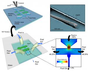
rice-u-cnt-uf-dvcFascinating research out of the Robinson, Kemere and Pasquali labs in Electrical & Computer Engineering and Chemistry Departments at Rice University, at Baylor College of Medicine and out of the Royer-Carfagni lab in Engineering at the University of Parma.  It was published recently in ACS NanoLetters and summarised in NewsMedical, and is focussed on the gentle introduction of microelectrodes for medical applications using microfluidics.  The team showed the insertion of carbon nanotube (CNT) electrodes into brain tissue surrogate (agarose gel) as well as rat brains, with subsequent observation of neural activity via these electrodes.  The ~30 µm CNT fibre is placed along the central channel in a glass/PDMS microfluidic device, and then carefully controlled sheath fluid flow is used to pull the wire down through a narrower ~100 µm-wide channel and into the tissue.  See impressive video showing microfluidic injection of CNT electrode into gel.

This technology is an excellent example of marrying microfluidics with nanotechnology, in this case for a medical application.  The microfluidic insertion of CNT electrodes in lieu of traditional approaches makes the process less damaging to surrounding brain tissue during insertion.  Neuro-sensing applications where gently introduced flexible CNT electrodes would be beneficial may include monitoring for epilepsy, repairing/patching connections for sight and hearing, as well as other areas of neuroscience research.

Open-source CAD for paper microfluidics

Open-source CAD for paper microfluidics

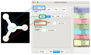
autopadResearchers from the Mace lab at Tufts University recently published an article in Scientific Reports highlighting their new open-source CAD software that is tailored for the design of paper microfluidic devices.  The software works across all OS platforms, appears fairly straightforward and user-friendly, and likely capable of covering most of the normal aspects of geometrical design for channel networks, based on examples presented.

The software is provided by the authors gratis in a laudable effort to reduce the costs associated with chip design, especially for resource-constrained environments such as schools and labs in developing countries.  The price for subscription or purchase of industry-standard CAD software can be prohibitive, easily reaching tens of thousands of dollars, and scaling with the number of users, etc.  Removing the cost of design SW breaks down a significant cost barrier to innovation for paper microfluidics, a technology that otherwise boasts very economical costs for consumable materials (paper) and fabrication (wax printing with commercially available printers).

Nanofluidic DNA sieving device from IBM

Nanofluidic DNA sieving device from IBM

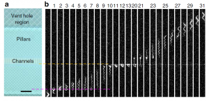
nf-dna-reptationFascinating research from the Qinghuang Lin lab at IBM Research in NY, published in Nature Communications and highlighted in Cytofluidix, shows an elaborate but potentially mass-producible microfluidic and nanofluidic lab-on-a-chip device that draws DNA molecules through arrays of nanometre-sized pillars.  The DNA reptation in these structure stretches the molecules out, as in gel-based electrophoretic separations of DNA, and may enable identification of biomarkers that indicate diseases.  The Cytofluidix summary page shows an engaging video of DNA migration through the pillar array.

The research team had to overcome sizable challenges associated with the nanofabrication of the different structures that act as the DNA sieving medium.  Researchers used a sacrificial silicon layer for the nanofluidic channels, and were able to overcome diffusion-limitted etching constraints by using temporary venting holes to accelerate the etching by 2 orders of magnitude.  This process improvement may be the key to making the device a manufacturable product instead of a clever academic exercise.

Origami switches for paper microfluidics

Origami switches for paper microfluidics

Research from Santosh Pandey‘s lab at Iowa State U, published in Lab on a Chip and highlighted in Cytofluidix, shows actuation of paper microfluidics switches/valves with effective, fast performance.  Several different configurations were demonstrated, including single-pole-single-throw (normally open and normally closed), single-pole-double-throw, by analogy to electrical switch terminology.  The switches appear to actuate in seconds, which is suitable for many paper microfluidics applications.  A video of the switches in action can be viewed on YouTube.

Several key advantages come to mind with this advance.  Paper microfluidics has simplicity, low cost manufacture and portability amongst its key attributes, but these are less credible if the devices also require expensive, power-intensive peripheral hardware for e.g. fluid movement, valve actuation, detector operation, communications, etc.  Implementation of this technology in a commercial product might reduce or eliminate the need for expensive pumps and valves (and accompanying electrical power consumption) for fluid flow and valving on a device.  This could enable very low cost analysis applications targeting harsh or remote environments.

Microfluidic nanoparticle analysis by Spectrodyne

Microfluidic nanoparticle analysis by Spectrodyne

spectradyne-uf-coulter-counter-deviceA recent article by Azo Materials featured a microfluidic nanoparticle analyser product from Spectrodyne.  The “nCS1” system appears to reap the benefits of precise small volume fluid manipulations in disposable microfluidic cartridges to eliminate sample cross-talk and any need for cleaning.

The system is an advanced Coulter counter, and has sufficiently high resolution to allow it to achieve superior measurements of particle polydispersity in a given sample.  Particle sizing is from 40-2000 nm for 10^5 to 10^12 particles/mL, with precision of better than +/- 3% on sizing and +/-10% on concentration.