Skin-on-chip system for modelling herpes simplex virus (HSV) infection

Skin-on-chip system for modelling herpes simplex virus (HSV) infection
Structure of skin-on-chip microfluidic vascular network and epidermis. (Image © Nature Communications/Jia Zhu et al.)

Impressive work out of the Zhu lab at the University of Washington’s Department of Laboratory Medicine & Pathology and Institute for Stem Cell & Regenerative Medicine appears to effectively mimic human skin by combining a microfluidic “blood vessel-like” vascular network and a stratified epidermis structure. The research was published in September in Nature Communications, and profiled by the UW’s Fred Hutch Cancer Centre.

The skin-on-chip device was constructed in several steps. First, a microfluidic network with 100 µm-wide channels was patterned via soft lithography/injection moulding in a collagen matrix with dermal fibroblasts. The channels were perfused with vascular endothelial cells that lined the surface and formed a vascular network. Second, at the outer surface of the collagen chip, the epidermis was constructed: epidermal keratinocytes were seeded for 3 days, and later exposed to air to differentiate into a multilayered epidermis. The graphic above shows diagrams and a confocal micrograph that illustrate the skin-on-chip device.

The growth of the seeded keratinocytes into a fully stratified epidermis over ~2 weeks was characterised with cross-sectional images and a variety of fluorescent dyes to show the growth of the layers. Comparisons of different fluorescently stained keratinocyte markers (K14 & K10), basement membrane markers (Col IV) and vimentin in the skin-on-chip model and in native skin showed the model to accurately mimic the natural structures in native skin.

Many experiments were performed to illustrate the abilities of their platform; two are showcased here. In one experiment, the effectiveness of herpes simplex virus 1 and 2 (HSV-1 and -2) infection was tracked at various stages of dermal growth, pinpointing dramatic decreases in infection rate with completed layers of dermal growth as days progressed. After just 1 day of exposure to air and differentiation of the dermal layer, the number of infected cells dropped from 67 to 16% for HSV-1, and from 100 to 36% for HSV-2. It’s no surprise that, as skin forms or heals, its ability to prevent infection improves, but accurately determining at what time, stage of growth and to what extent this happens via a real model is powerful indeed.
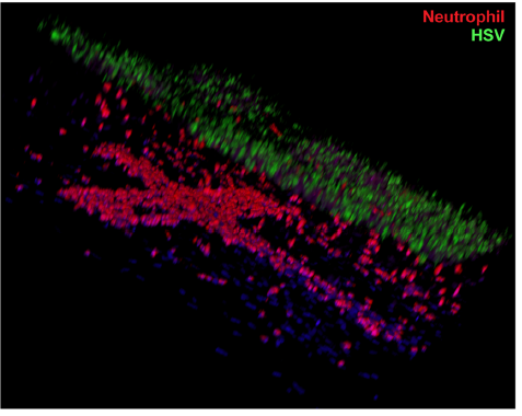
White blood cell neutrophils (red) migrating up to HSV- infected skin (green) in an on-chip immune response. (Image © Nature Communications/Jia Zhu et al.)

In another experiment, the authors showed that neutrophils (the most abundant type of the white blood cells used in our body’s immune response) would adhere to the walls within minutes of perfusion through the microfluidic vascular network for a chip that was infected with HSV, vs. essentially no adhesion in a control chip. After hours, the neutrophils continued to accumulate, transmigrated through the endothelium and entered into the layered dermis on the chip surface. A confocal microscopy image, reproduced at right, shows the presence of red-stained neutrophils in the cross-shaped microfluidic vasculature below, and migrating upwards towards the HSV-infected skin layers above, stained green.

The effectiveness and utility of this skin-on-chip model system to monitor immune responses to disease is obvious. The authors discuss an array of investigations that can be undertaken to better understand the chemistry of immune responses. If we consider the 3-D spatial information content provided by confocal microscope imagery in the context of their stratified, vascular-epidermal microfluidic system, it seems like the sky is the limit for recreating immune and other skin-based biological/biochemical processes, and perhaps with other tissue types as well. There is a good reason this paper was accepted by Nature Communications. 🙂

Microfluidic fabrication of hydrogels that consume environmental pollutants

New work from Patrick Doyle’s group at MIT, just published in ACS’ Applied Polymer Materials and highlighted in MIT News, shows the simple microfluidic manufacture of hydrogel particles that can be used to remove hydrophobic (non-polar, water ‘fearing’) micropollutants from water. Their hydrogel ‘filtres’ show enhanced performance compared to activated carbon (AC); in addition, and critically, they can easily be regenerated.

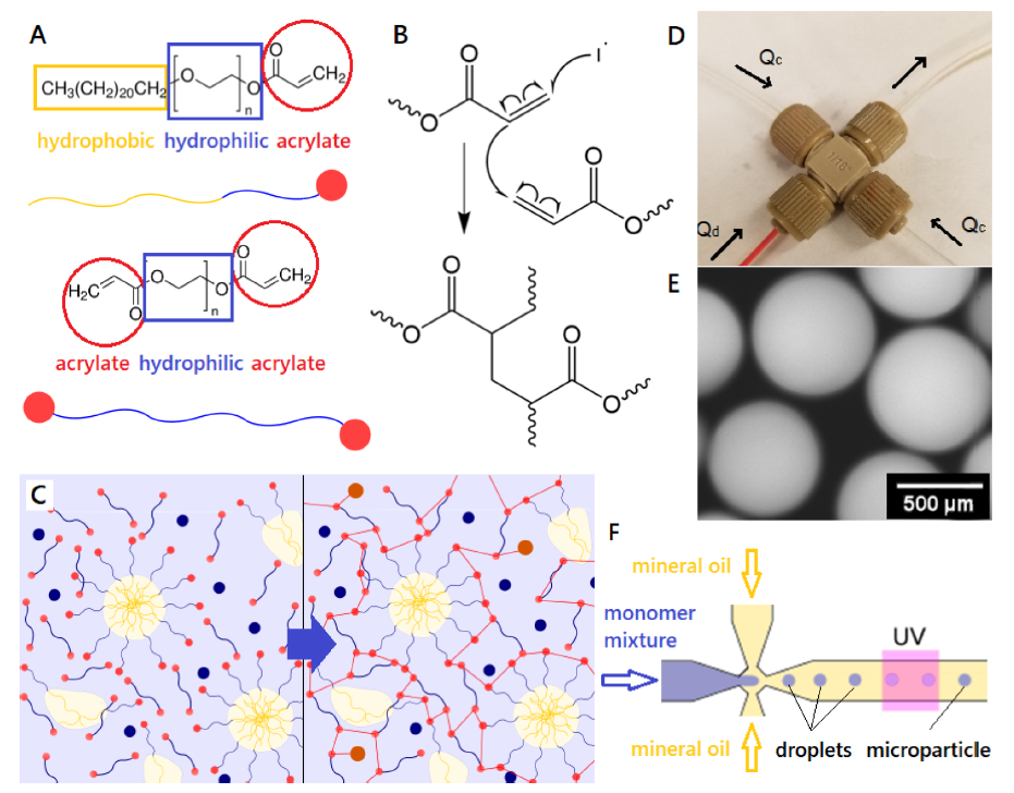
Microfluidic synthesis of micelle-based hydrogel particles. (Image © American Chemical Society/Applied Polymer Materials)

The hydrogel particles are made from acrylate-based micelles that are covalently bound into a cross-linked acrylate gel. The diagram above from their paper illustrates the nature of the micelle monomers and cross-linkers (A); the polymerisation reaction (B – I’ll pretend I understand exactly what’s going on, there …;-); the solution (left) before polymerisation, with free micelles spontaneously formed, and (right) after (UV) polymerisation, with the micelles fixed in the gel polymer (C); the microfluidic cross used for droplet/particle formation (D); a micrograph of the 500 µm gel particles (E); and a diagram showing the formation of the droplets in the microfluidic reactor that polymerise under UV light to form the gel particles.

The microfluidic reactor in this case is simply made from off-the-shelf components: a microcross such as from Idex (150 µm through-hole) and 1/16″ OD tubing. For R&D, this is an ideal set-up: fairly cheap with parts delivered in days and set up in minutes. For manufacturing, it’s only a small leap to replicate this structure in polymer or glass and set multiple microfluidic synthesis networks up together in parallel to reduce the cost contribution of the microfluidic reactor.

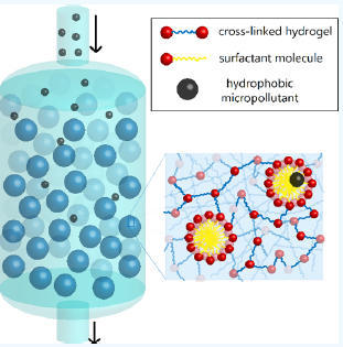
Hydrogel column used to clean water. (Image © American Chemical Society/Applied Polymer Materials)

To remove the pollutants from a water sample, water is passed through a column filled with this hydrogel, and the hydrophobic pollutants partition into the hydrophobic micelle centres, analogous to reverse-phase chromatography, with the pollutants being highly retained in the stationary phase. Once the hydrogel is saturated with pollutants, it can be regenerated with an ethanol flush, purging the column of bound pollutants; the authors claim it will maintain performance over years of such regenerative cycles. Different micelle monomer hydrophobic tails can be used to optimise the affinity and partitioning coefficient for a variety of pollutant targets.

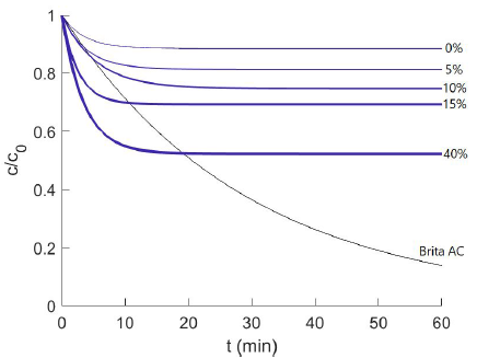
Uptake of 2-napthol by F127DA hydrogel at different concentrations, compared to activated carbon. (Image © American Chemical Society/Applied Polymer Materials)

The performance of the hydrogels stacks up very nicely against activated carbon filtres which are the current gold standard in many applications. Doyle’s group measured the performance of a variety of different micelle compounds in their hydrogels against each other and activated carbon for their micropollutant model compound, 2-napthol (used in the manufacture of dyes, fungicides, insecticides, pharmaceuticals and perfumes; carries the H400 classification of “very toxic to aquatic life”). The graph at right plots pollutant removal over time for a particular micelle formulation, F127DA. The ordinate is c/c(o), or the concentration of pollutant left in the test solution vs. initial concentration; the faster and greater this drops, the better the performance. The four different exponential decay curves (blue) correspond to different quantities of micelle surfactants bound in the hydrogel: 0 to 40% surfactant concentration in the gel; the grey curve is for Brita activated carbon. The plot shows that higher percent quantities of micelles bound in the hydrogel lead to pollutant removal in greater quantities and at faster rates. On time scales under 10 minutes, typical for water treatment contact times, both the 15 and 40% F127DA formulations handily outperform the activated carbon, although longer timescales favour the AC.

The authors highlight the fact that their hydrogels can be regenerated with ethanol, using between one thousandth to one billionth the volume of EtOH vs. water treated, and show good performance for cycles subsequent to regeneration. They also note the very energy-intensive process needed for regenerating AC filtres. While it seems they have effectively shown good performance from their hydrogels for both filtration and re-usability, what is missing in the comparison, in my view, is a clearer contrast of economic and environmental drivers. For example, potential costs/year for filtre replacement, ethanol/energy consumption, releases to the environment, etc. That is arguably the product developer’s job, and perhaps this paper’s performance will pique the interest of some entrepreneurs, hopefully with well-heeled partners.

Simplicity through capillarity: automatic microfluidic analyses

New research from David Juncker’s lab at McGill University’s Biomedical Engineering Department show’s impressive progress along the path to sophisticated diagnostics or analyses performed on low-cost microfluidic devices in resource-limited environments. Their work was just published last month in Nature and also profiled in an article in McGill’s Health e-News.

Juncker’s group excels at capillarity-driven microfluidic devices, and they wrote an excellent critical review paper in Lab on a Chip a few years ago on ‘capillaric circuits’. It provides a full description of the physical chemistry of Laplace pressure and surface chemistry that underpin the capillarity-driven microfluidic operations used in these devices. While the full depth of the chemistry and physics is beyond the scope of this blog article (and the author’s RAM capacity ;-), they may be explored separately in a future post. Suffice it to say that, while somewhat involved, the capillarity-based pumping, valving and timing technology is well understood and and fairly well developed by this group, amongst others.

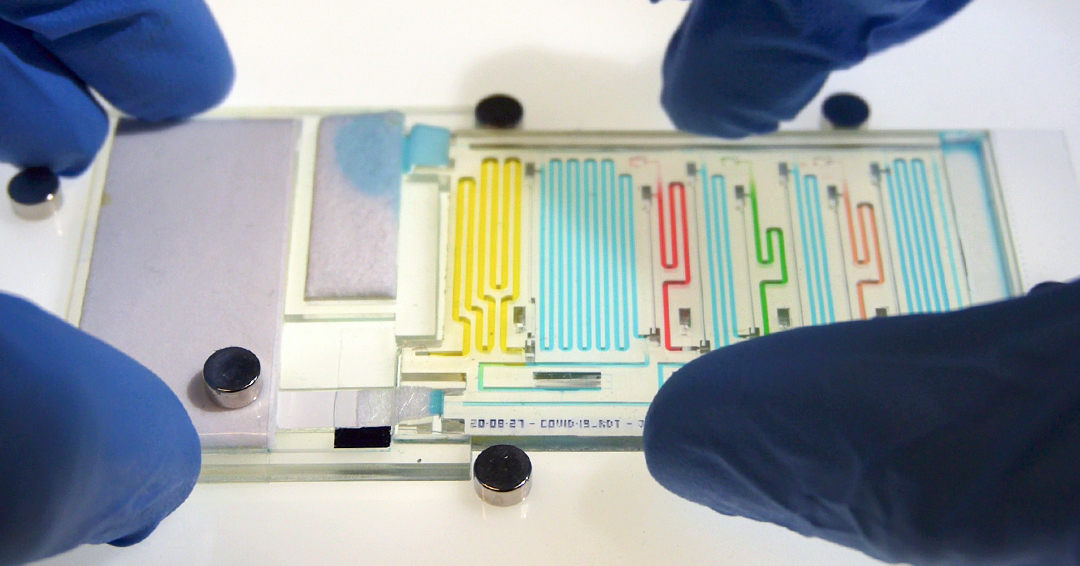
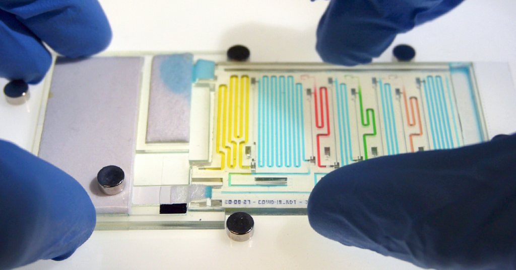
‘Microfluidic Chain Reaction’ device for COVID-19 detection (image © McGill University Health e-News)

In this work, they are moving from the ‘mere’ sequential delivery of a few liquids at a channel intersection to fully configured, multi-step analyses using hundreds of sequential pre-programmed liquid manipulations. A first chip enables sequential delivery of 300 sample aliquots, while the second, shown above, performs the 8-step ELISA detection of COVID-19 antigen found in a patient’s saliva sample . Some impressive videos showing the performance of the 300 aliquot chip and the COVID-19 diagnostic chip can be found in the McGill article.

For someone like myself working in the field of microfluidics, perhaps the most thought-provoking aspect of this article is the potential for the technology to dramatically simplify the operation of fairly complex microfluidic devices. Being able to programme sequential volumetric flows with precise timing with a virtually unrestricted number of on-chip steps, all under autonomous operation, has far-reaching implications. It means that, after addition of sample(s), the device will simply start its protocol, with no interaction needed from an instrument or human operator. Using existing, fully developed on-board technologies such as lyophilised reagents and mixing procedures together with simple visible bands for detection such as with COVID-19 rapid tests, the door to many other more complex medical diagnostics, industrial or environmental assays is now open.

Development of products to meet these application needs will no doubt carry challenges, but at the end is a cheaply manufactured, complex device that can be used at the point of care or need; this represents a huge foundation on which to build any number of enabling, disruptive products.